
High quality studies need valuable histological analysis. One room in EMIS is dedicated to histopathology, in which a microscope (Nikon, Eclipse E800), and a whole slide imaging system (virtual microscopy or scanner Coolscan V, Nikon) are available to researchers.
This scanner allows performing digital images in high resolution leading to representative and reproducible analyses.
Histological interpretation may be performed either by means of the platform, either by the researchers themselves.
Furthermore, innovative and efficient technics such as multiphoton microscopy are available in partnership with XLim research team.
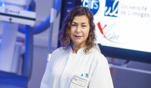 Professor Catherine YARDIN


©2021 Graphik Studio • All Rights Reserved • Legal notice